
Elbow Dysplasia in Dogs: Front-Leg Limping, Diagnosis, Treatment Options and Costs
By Cal Hewitt · Published , updated
Health
Elbow dysplasia in dogs is a group of developmental problems in the elbow joint. It can cause front-leg lameness, stiffness, pain, reduced elbow movement and arthritis, often beginning while a dog is growing. The main forms are medial coronoid disease, ununited anconeal process, osteochondrosis (also called osteochondritis dissecans) and elbow incongruity. A vet may use an orthopedic exam and X-rays, with CT or arthroscopy when standard images don't show the problem clearly. Treatment depends on the lesion and how much arthritis is present, and it can include weight and activity management, vet-directed pain control, rehabilitation or surgery. A limp that keeps coming back should be examined, and a dog that suddenly can't use the leg should be seen today.
Key takeaways
Tap a card to bring it forward.
- Elbow dysplasia is an umbrella term for several developmental elbow problems, not one single disease.
- Front-leg lameness that worsens after exercise, stiffness after rest or a limp that keeps returning is a reason to book a vet visit soon.
- Sudden inability to use the leg, severe pain, a deformed limb or a fall or collision means a same-day vet visit.
- Normal-looking X-rays do not always rule it out, and both elbows are often involved.
- Surgery cannot cure it or reverse arthritis that is already there, and many dogs do well with combined treatment.
- Lean weight, controlled exercise and secure footing help every dog with it, and you should never give a human pain reliever.
My dog is limping on a front leg: is this a visit soon or a visit today?
Watching a young dog limp after a good run is worrying, and you want to know how fast to move. This is the sorting I'd use; your vet has the final say.
Book a vet appointment soon if you see any of these: a front-leg limp that keeps coming back, a limp that gets worse after exercise, stiffness after rest, pain when the elbow bends or straightens, less willingness to walk or play, a swollen or thickened-looking elbow, or repeated episodes in a growing dog. A limp that improves and then returns still counts. Note when it began, and if it is safe, film a short video of your dog walking to show the vet.
Go to a vet the same day for any of these: your dog suddenly can't bear weight, the pain is severe or getting worse, the limb looks deformed or is badly swollen, there was a fall or collision, or your dog can't stand or walk normally. The same goes for a limp with marked tiredness, collapse or trouble breathing. You can find a clinic in the DogServ veterinarian directory or, after hours, the emergency vet directory.
Please don't reach for your own medicine cabinet; human pain relievers can poison dogs, and any pain medicine should be the one and the amount your vet gives you. Until the exam, keep walks short and calm on a leash, and skip fetch, jumping and rough play. If you aren't sure the problem is in the elbow, my guide to why a dog is limping covers the wider set of causes.
What elbow dysplasia means: the four forms in plain words
The word "dysplasia" sounds like one diagnosis, but the American College of Veterinary Surgeons (ACVS) and the Merck Veterinary Manual both describe it as a group of developmental abnormalities of the elbow. Your vet or surgeon may name one or more of these:
- Medial coronoid disease, or fragmented coronoid process. Part of a small bony knob on the ulna (a forearm bone) is diseased or has broken off. The fragment can be bone, cartilage or both.
- Ununited anconeal process. A piece of the ulna that should fuse during growth stays separate or loose.
- Osteochondrosis, or osteochondritis dissecans (OCD). The cartilage and the bone under it develop abnormally, and sometimes a loose flap or fragment forms.
- Elbow incongruity. The radius, ulna and humerus don't fit together the way they should, so the load spreads unevenly and can contribute to the other problems.
All of these can damage cartilage and lead to osteoarthritis over time, and a dog can have more than one at once.
This is a front-leg joint problem. Hip dysplasia affects the back-leg hip joint. Both are developmental and can lead to arthritis, but the joints, signs, imaging and procedures differ. I cover the other one in my guide to hip dysplasia in dogs.
The signs, the usual age and why a dog can have it without limping all the time
Common signs include front-leg lameness that is often worse after exercise, a limp that recurs instead of clearing up fully with rest, stiffness after resting, pain when the elbow is bent or extended, reduced range of motion, joint fluid or swelling, and reluctance to exercise. Some dogs hold the elbow away from the body, and long-standing cases can bring muscle loss or a grating feeling in the joint.
Age varies. Merck places typical signs at roughly 4 to 10 months in young, fast-growing medium, large and giant dogs. ACVS says signs commonly appear from about 5 months, and that some dogs aren't diagnosed until 4 to 6 years old. So, an adult dog with a new front-leg limp is still a dog worth having examined for this.
Both elbows are often involved. ACVS reports that both may be affected in up to 80% of patients, so a vet may want to look at both. The Orthopedic Foundation for Animals (OFA) adds that the changes seen on X-rays and visible lameness don't line up directly. A dog can have elbow disease on film without limping every day. OFA also says a Grade I elbow should not be assumed to progress to Grade II or III.
How the vet confirms it: the exam, X-rays, CT and arthroscopy
Diagnosis starts with your dog's history, a look at the walk and a hands-on orthopedic exam. The vet checks pain when the elbow bends and straightens, range of motion, swelling and muscle loss, and whether one leg or both are involved.
X-rays. Radiographs can show arthritis changes, bone spurs, an ununited anconeal process or a mineralized fragment. OFA's screening uses a view with the elbow fully flexed. X-rays can miss cartilage-only or subtle coronoid lesions.
CT. A CT scan gives cross-section and three-dimensional images, and it is especially useful for subtle bone lesions, incongruity and medial coronoid disease. Both elbows are commonly scanned.
Arthroscopy. A camera and small instruments go into the joint, so the surgeon can see the cartilage and lesions directly. It can also allow treatment during the same procedure.
Why does this matter? A 2009 case series followed 16 dogs with front-limb lameness from the elbow and no changes on X-rays. CT or scintigraphy suggested a problem, and arthroscopy confirmed medial coronoid disease in every abnormal elbow. So, if your dog keeps limping and the X-rays look clean, ask your vet whether CT or a specialist opinion is the next step.
What causes it: growth, inherited risk, weight and diet
ACVS describes elbow dysplasia as multifactorial. Suspected contributors include inherited risk, abnormal cartilage development, trauma, diet and growth-related disturbances. Merck identifies young, rapidly growing medium, large and giant dogs as the typical patients. A breed on its own doesn't prove a diagnosis; it is a risk factor, and the exam and imaging decide.
Food deserves a careful word. The COAST Development Group consensus says nutrient deficiency or excess, an imbalance of calcium and phosphorus, too many calories and overly rapid growth can contribute to skeletal and joint problems, and that puppies need nutrition suited to growth. That does not show that one food caused one dog's disease. If you have a large-breed puppy, ask your vet which growth-appropriate diet and body condition to aim for, and feed the amount the vet gives you.
Surgery or not: what each option involves and what to expect afterward
Not every dog with elbow dysplasia needs surgery. ACVS allows medical management when the disease is very mild, or when the joint is so damaged that surgery is unlikely to help. The decision depends on the lesion, your dog's age, the degree of arthritis, pain and what the imaging shows.
Surgery may include:
- Arthroscopic removal of coronoid fragments or loose cartilage, and arthroscopic treatment of OCD lesions.
- Fixing or removing an ununited anconeal process.
- An ulnar osteotomy (a cut in the ulna) or other procedures to address incongruity or move the load.
- Open surgery where arthroscopy isn't suitable, and joint replacement or other salvage procedures in severe, end-stage disease.
Now the honest part. ACVS states that surgery cannot cure elbow dysplasia or reverse established arthritis, and that arthritis may keep progressing even when comfort improves. ACVS also reports that, on average, 85% of cases show some improvement in lameness and comfort with treatment, and that patients can have a good long-term outcome through combined surgical and medical management; it says the condition "cannot be cured but it can be well managed." Evidence on whether arthroscopy beats conservative care for medial coronoid disease is thin. A 2025 systematic review of three studies found weak evidence, small groups and no clear answer that arthroscopy is better. So I can't tell you surgery always gives a better long-term result, and a good surgeon won't promise it either.
A 2023 case series of 23 dogs with OCD at the end of the humerus, all treated arthroscopically after CT, found that 90% also had medial coronoid disease. That is a selected group and not a head-to-head trial.
Daily life with it: lean weight, controlled exercise, secure footing, rehabilitation and medicine classes
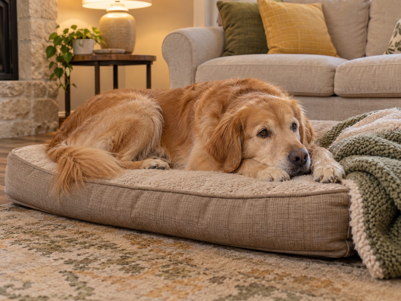
Home care carries a lot of the work, whatever the treatment. The AAHA pain management guidelines and the COAST consensus both put weight, nutrition, appropriate exercise, rehabilitation, changes at home and regular reassessment at the foundation of joint care.
- Keep your dog lean. Ask your vet to assess your dog's body condition and say how much, if any, weight should come off. Extra weight loads sore elbows with every step.
- Use regular, controlled exercise. Steady leash walks fit your dog's ability better than weekend sprints. Avoid sudden loading, repeated jumping, high-impact play and sharp turns, especially in an unconditioned dog. My guide to how much exercise a dog needs covers the general principles; your vet sets the elbow-specific limits.
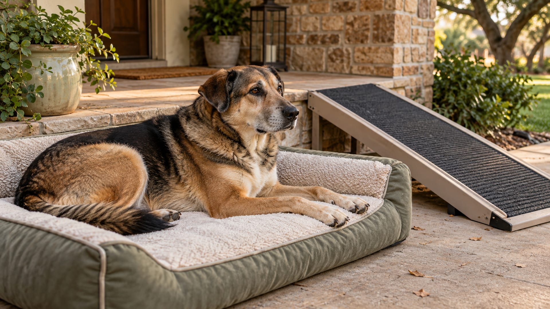
- Make the floor safe. Carpet runners or other secure footing help on slippery floors. Ramps or steps can replace jumps onto the couch or into the car.
- Give a good place to rest. Put comfortable bedding where your dog can reach it without stairs.
- Use rehabilitation if you can. A veterinary rehabilitation professional can set up exercises that fit your dog.
- Watch and report. Notice how your dog walks, gets up, handles stairs and plays, and whether appetite or mood changes. Tell your vet if comfort or mobility gets worse.
Your vet may use medicine classes such as NSAIDs (non-steroidal anti-inflammatory drugs), and may consider joint-support products, including nutraceuticals or polysulfated glycosaminoglycans. AAHA reports low evidence for most nutraceuticals, with stronger support for omega-3 fatty acids than for other supplements, so a supplement is an extra and not a stand-in for weight control, rehabilitation or vet pain care. My page on glucosamine for dogs covers what it can and can't do. Use only the product and amount your vet gives you, and follow any monitoring they ask for.
For comfort as your dog gets older, see my guide to senior dog care.
What it costs
These US estimates come from VetCostGuide, published February 10, 2026 and updated June 6, 2026. They rest on public data and industry surveys and are not an official fee schedule, so treat them as a rough range; costs vary by region and hospital.
Tap or hover a row to highlight it.
| Item | Estimate |
|---|---|
| ItemX-rays plus CT | Estimate$500 to $1,500 |
| ItemArthroscopy | Estimate$1,500 to $4,000 per elbow |
| ItemOpen surgery or osteotomy | Estimate$2,500 to $5,000 per elbow |
| ItemRehabilitation | Estimate$500 to $2,000 |
| ItemLong-term arthritis management | Estimate$100 to $300 per month |
| ItemEstimated lifetime cost | Estimate$5,000 to more than $20,000 |
Bills run higher when both elbows are affected, or when referral, anesthesia, hospitalization, repeat imaging, complications or joint replacement come into it. Ask the treating hospital for a written, itemized estimate covering the consultation, imaging, anesthesia, surgery, medicines, hospitalization, rechecks and rehabilitation. If a diagnosis is already on your dog's record, ask an insurer how it treats pre-existing conditions; my guide to pet insurance for dogs covers coverage and exclusions.
Elbow screening (OFA) and inherited risk
OFA elbow screening uses X-rays of both elbows, and a final normal certification isn't issued until the dog is 24 months old. OFA grades abnormal elbows by the secondary arthritis it sees:
- Grade I: bone spurs (osteophytes) under 2 mm.
- Grade II: bone spurs from 2 to 5 mm, with other changes.
- Grade III: bone spurs over 5 mm, with well-developed degenerative joint disease.
Inherited risk is supported, though the condition is multifactorial. OFA says signs alone aren't a reliable basis for breeding decisions and recommends X-ray information for dogs and their siblings in at-risk breeding programs. Selective breeding based on screening can lower inherited risk, but it doesn't guarantee that every puppy will be unaffected.
Questions dog owners ask about elbow dysplasia
These are the questions people search most, answered briefly.
How long can a dog live with elbow dysplasia?
ACVS says patients can have a good long-term outcome with combined surgical and medical management. The arthritis can keep progressing, so the goal is comfort and movement over the years, with regular rechecks.
How do you treat elbow dysplasia in dogs?
Treatment depends on the lesion, age and arthritis. It can include weight and activity management, vet-directed pain medicine, rehabilitation and surgery such as arthroscopy, open procedures or, in severe cases, joint replacement.
Can a dog live with elbow dysplasia without surgery?
Yes, in some cases. ACVS allows medical management for very mild disease or when surgery is unlikely to help a badly damaged joint. Ask your vet which path fits your dog.
What does elbow dysplasia in dogs look like?
Usually a front-leg limp that is worse after exercise, stiffness after rest, a reluctance to walk or play, and sometimes a swollen elbow or an elbow held away from the body. A dog with both elbows affected may refuse to finish a walk.
Your next step
If your dog is limping on a front leg, start with a note of when it began and a short video of the walk, and book an exam soon. Go the same day if your dog can't use the leg, is in severe pain or was hurt. At the visit, ask whether both elbows should be checked, whether X-rays are enough or CT makes sense, and ask for a written estimate. Find a clinic in the DogServ veterinarian directory.
Sources
- American Animal Hospital Association: AAHA pain management guidelines
- American College of Veterinary Surgeons: American College of Veterinary Surgeons (ACVS)
- Merck Veterinary Manual: Merck Veterinary Manual
- ofa.org: Orthopedic Foundation for Animals (OFA)
- PubMed: 2009 case series
- PubMed Central: 2023 case series
- PubMed Central: 2025 systematic review
- PubMed Central: COAST Development Group consensus